Elbow Injury Tennis Elbow

Question:  I don’t play tennis; how can I have tennis elbow?

Answer:  Easily, lateral epicondylitis, a.k.a. tennis elbow has little to do with tennis and lots to do with constantly using your arm in a repetitive motion.

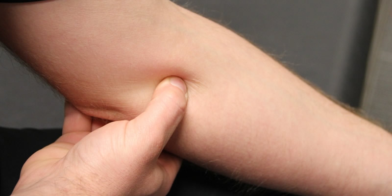
Epicondyles are the bony bumps on the inside and outside of your elbow. Epicondylitis is the inflammation of these. 95% of people suffering from this do not play tennis, but this is a common sports related injury. This can be a result of any sport where your arm is in a similar position or even work related such as carpentry, plumbing or constant use of a computer keyboard. Even musicians are vulnerable!

After repeated use, the tendons at the elbow end of the extensor carpi radialis brevis (ECRB) muscle may develop small tears. This leads to inflammation and stress on the rest of your arm. It is a form of tendonitis.  This can be a result of any activity that requires repetitive arm, wrist, or elbow movement. The extensor tendons are used when extending your wrist and fingers. The flexor tendons are needed for closing wrist and fingers. Both flexor and extensors are used in thousands of tasks, and often handle very high levels of stress.

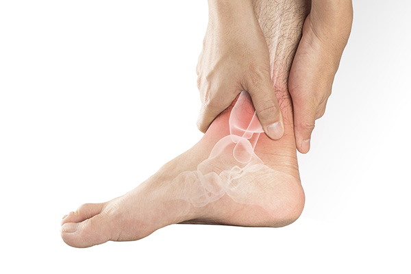
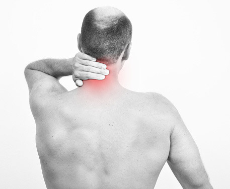
Inflammation is a normal part of the healing process and the body’s natural response to an immediate injury. Swelling, discomfort, the sensation of heat, redness, and loss of function are the key symptoms which should warn you something is not right in your world. Tenderness and stiffness are also red flags, and when the pain is severe enough, it may radiate down the arm, into the hand and fingers.

Approximately 3% of the population experiences this injury, typically occurring in men and women between the ages of 40 and 50. Your daily activity is a big determining factor in your likeliness to be affected.  Office workers, secretaries and Health Care Workers are especially prone.

Self-diagnosis is a dangerous thing as there may be more than one potential issue with the same symptoms.

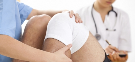
As with any injury or ailment, the first step is to visit your physician. Dr. Marshall Allegra spends quality time speaking with each patient at his Hazlet office to understand their unique condition based on symptoms and life-style. He has over 25 years’ experience diagnosing pain and prescribing the most appropriate treatment, exploring all non-surgical possibilities first. Common remedies and treatments Dr. Allegra may suggest:

  • Cold therapy treatments should be in the earliest stages of an injury only as this will actually impede the recovery because cold restricts the vessels – reducing the blood flow to the area
  • Anti-inflammatory medication may relieve the pain
  • Limit activities for a few days or possibly weeks until the inflammation subsides. If possible, avoid the repetitive movement.
  • A brace may help stabilize the area and limit movement for you
  • Supervised strengthening and stretching exercises may help, but be sure to do the correct ones in the correct position or you may further injure yourself. A physical therapist can work out a routine for you to safely improve your symptoms.
  • Acupuncture
  • Steroid injections directly into the inflamed area target the inflammation.
  • Surgery when all else fails.

It is essential to allow all injuries to heal completely before returning to your daily activities.  You want to avoid the build-up of scar tissues or having on-going issues. Be sure to follow up with Dr. Allegra before returning to your routine.

Contact Dr. Marshall P. Allegra

879 Poole Avenue, Hazlet New Jersey, 07730
Phone: (732) 888-8388

mallegra879@yahoo.com

What are the most common orthopedic surgeries?

Orthopedic surgery is a surgical procedure performed on the musculoskeletal system for the correction of injury or other conditions. The musculoskeletal system is made up of bones, joints, and soft tissues such as muscles, ligaments, and tendons. Any part of the musculoskeletal system can become impaired through injury/accident or caused by other degenerative conditions.


Orthopedic surgery can performed through traditional open surgery, or arthroscopically. Arthroscopy is a surgical procedure on a joint that is minimally invasive as treatment is provided using an arthroscope inserted into the joint through a small incision.

Most orthopedic surgeries are performed on the ankle, knee, hip, wrist, elbow, shoulder or spine. The most common procedures include:

  1. Soft tissue repair, for torn ligaments and tendons.
  2. Joint replacement, in which a damaged joint is replaced with a prosthesis.
  3. Revision joint surgery, in which a preexistent implant is changed with a new one.
  4. Bone fracture repair
  5. Debridement, during which damaged soft tissues or bones are removed.
  6. Fusion of bones, in which bones are fused with grafts.
  7. Spine fusion, during which the spinal bones (vertebrae) are joined together.
  8. Osteotomy, aimed at correcting bone deformities.


Dr. Marshall P. Allegra is a board-certified orthopedic surgeon with over 23 years experience treating patients in Monmouth County. Depending on the type of condition or injury, Dr. Allegra may recommend fixing a fracture with rods, plates, and screws that will hold the fracture together during healing. Contact the offices today for answers to any questions you may have or to schedule an appointment.

Arthroscopic Surgery in Hazlet NJ

Joint problems are common simply due to the amount of pressure we put on our bodies. Our joints are constantly flexing and bending as we go about our daily life which can cause major wear and tear to our bodies over time. Arthroscopic surgery uses a camera to look at the damage to your joints to determine what (if anything) needs to be done. An arthroscope can technically be used anywhere in the body but is generally needed for the knees, ankles, wrists, hips, and shoulders.

The Basics of Arthroscopy

Arthroscopy is used to either diagnose or help treat general inflammation or trauma. It’s generally an outpatient procedure, but you will be given some type of anesthetic. Depending on the severity of the injury and the location of the damaged joint, it may be either general or local anesthesia. The doctor will make a tiny cut in your skin before inserting an instrument into your body (about the size of a standard pencil.) The doctor will fill the joint with fluid to make it easier to fit the tiny camera inside.

Next Steps

The arthroscope is used to let the doctor look inside the joint to see if you need arthroscopic surgery. Should you need the surgery, your doctor will need to manipulate your bones to fix the joints. This can be done without making large incisions, but rather using precise instruments to shave or cut the bone. You may not even need stitches! If for any reason you do need traditional surgery, this can be done in the same procedure as the arthroscopic surgery. Dr. Allegra at Allegra Orthopedics serves the people of Hazlet, NJ and understands the ins and outs of joints. He’s here to help you increase your flexibility and range of motion while simultaneously decreasing any discomfort!

What Is a Hairline Fracture?

We often hear the term hairline fracture, but do you know what the term means?

Orthopedic physicians describe hairline or stress fractures as tiny cracks that develop on bones in the foot or lower leg. A common injury in runners, gymnasts, and dancers, anyone can develop a hairline fracture through repetitive jumping or running. Individuals with osteoporosis also are at risk for hairline fractures.

The bones of the foot and leg absorb a lot of stress during running and jumping. Common sites for hairline fracture include the metatarsal bones – the five long and narrow bones connecting the mid-foot to the toes – as well as the heel, ankle bones, and the navicular, the bone on the top of the mid-foot.

What are the symptoms of a hairline fracture?

The most common symptom is pain that gradually worsens over time, especially if you don’t stop weight-bearing activity. Other symptoms include swelling, tenderness, and bruising.

What causes a hairline fracture?

An overuse or repetitive activity, or an increase in either the duration or frequency of activity, can result in a hairline fracture. Even regular runners who suddenly increase either their distance, such as someone training for a marathon, or the number of times per week they run, can cause a hairline fracture.

Another cause of a hairline fracture is changing the type of exercise normally performed.

Bones adapt to increased forces through various activities, where new bones form to replace old bone. This process is called remodeling. When the breakdown of bone happens more rapidly than new bone can form, the likelihood of developing a hairline fracture increases.

Who’s most at risk for developing a hairline fracture?

There are several risk factors that increase the chances of developing a hairline fracture:

  • Certain sports: Participants in high-impact sports such as track and field, basketball, tennis, dance, ballet, long-distance runners, and gymnastics, increase their chances of getting a hairline fracture.
  • Gender: Post-menopausal women have an increased risk of hairline fractures. Female athletes may be at a greater risk because of a condition called the “female athlete triad.” This is where extreme dieting and exercise may result in eating disorders, menstrual dysfunction, and premature osteoporosis.
  • Foot problems: Poor footwear can cause injuries, so can high arches, rigid arches, or flat feet.
  • Weakened bones: People with osteoporosis, or anyone taking medications that affect bone density and strength, can develop hairline fractures even when performing normal activities.
  • Previous hairline fractures: Having one hairline fracture increases your chances of having another.
  • Lack of nutrients:Lack of vitamin D or calcium can make bones more susceptible to fracture. People with eating disorders are at risk for this reason. There can be a greater risk of hairline fractures during the winter months when diminished sunlight decreases the body’s absorption of vitamin D, compounded by an increased risk of slips and trips on icy surfaces.
  • Improper technique: Blisters, bunions, and tendonitis can affect how you run, altering which bones are impacted by certain activities.
  • Changes in surface: Changes in playing surfaces can cause undue stress to the bones of the feet and legs. For example, tennis player who move from a grass court to a hard court may develop injuries.

How is a hairline fracture diagnosed?

If you believe you have a hairline fracture, it’s important to seek treatment from an orthopedic physician as soon as possible.

Your doctor will perform a physical exam and review your medical history and general health, including diet, medications, and other risk factors. Diagnostic tools may include an MRI, X-ray, or bone scan.

How are hairline fractures treated?

If you suspect you have a hairline fracture, follow the RICE method: rest, ice, compression and elevation. An over-the-counter, non-steroidal drug can help with pain and swelling.

It’s important to seek further treatment if the pain becomes severe or doesn’t get better with rest. The treatment your orthopedic physician recommends will depend on both the severity and location of your injury.

Can other conditions develop if hairline fractures aren’t treated?

Ignoring the pain caused by a hairline fracture can result in the bone breaking completely. Complete breaks will take longer to heal and involve more complicated treatments. It’s important to seek advice from an orthopedic physician and treat a hairline fracture as soon as possible.

Medical treatments

Your doctor may recommend that you use crutches to keep weight off an injured foot or leg. You can also wear protective footwear or a cast.

Because it usually takes up to six to eight weeks to completely heal from a hairline fracture, it’s important to modify your activities during that time. Cycling and swimming are great alternatives to more high-impact exercises.

In some cases, a hairline fracture may require surgery. The addition of pins or screws can help hold bones together during the healing process.

What’s the outlook for someone with a hairline fracture?

It’s important to avoid high-impact activities during the healing process. Returning to high-impact activities — especially the activity that caused the injury in the first place — will delay healing and increase the risk of a complete fracture in the bone.

Even after the hairline fracture is healed, it’s important to gradually return to exercise.

In rare instances, hairline fractures don’t heal properly. This result is chronic, long-term pain. Talk with a board-certified orthopedic physician soon after the injury occurs to prevent pain and worsening damage to the bone.

Men and Osteoporosis

Osteoporosis is a disease that causes the skeleton to weaken and the bones to break. While most people think of osteoporosis as a woman’s disease, it poses a significant threat to millions of men in the United States.
There are many lifestyle habits that put men at increased risk of osteoporosis, yet few men recognize the disease as a significant threat to their mobility and independence.
Osteoporosis is called a “silent disease” because it progresses without symptoms until a fracture occurs. It develops less often in men than in women because men have larger skeletons, their bone loss starts later and progresses more slowly, and they have no period of rapid hormonal change and bone loss. However, in the past few years the problem of osteoporosis in men has become more recognized, particularly in light of estimates that the number of men above the age of 70 will continue to increase as life expectancy continues to rise.
Bone is constantly changing. Old bone is removed and replaced by new bone. During childhood, more bone is produced than removed, so the skeleton grows in both size and strength. For most people, bone mass peaks during the third decade of life. By this age, men typically have accumulated more bone mass than women. After this point, the amount of bone in the skeleton typically begins to decline slowly as removal of old bone exceeds formation of new bone.

Men in their fifties do not experience the rapid loss of bone mass that women do in the years following menopause. But by age 65 or 70, men and women are losing bone mass at the same rate, and the absorption of calcium, an essential nutrient for bone health throughout life, decreases in both sexes. Excessive bone loss causes bone to become fragile and more likely to fracture.
Fractures resulting from osteoporosis most commonly occur in the hip, spine, and wrist, and can be permanently disabling. Hip fractures are especially dangerous. Perhaps because such fractures tend to occur at older ages in men than in women, men who sustain hip fractures are more likely than women to die from complications.
In addition to age-related bone loss, there are many lifestyle behaviors, diseases and medications that can hasten bone loss in men. These include:
• Chronic diseases that affect the kidneys, lungs, stomach, and intestines, or alter hormone level
• Regular use of certain medications, such as glucocorticoids
• Undiagnosed low levels of the sex hormone testosterone
• Unhealthy lifestyle habits: smoking, excessive alcohol use, low calcium intake, and inadequate physical exercise
• Age. The older you are, the greater your risk
• Race. Caucasian men appear to be at particularly high risk, but all men can develop this disease.
Osteoporosis can be effectively treated if it is detected before significant bone loss has occurred. An orthopedic physician can order a medical workup to diagnose osteoporosis, including a complete medical history, x-rays, and urine and blood tests. The doctor may also order a bone mineral density test to determine your risk for fractures (broken bones), and measure your response to osteoporosis treatment.
In men, osteoporosis is often not diagnosed until a fracture occurs or a man complains of back pain and sees his doctor. This makes it especially important for men to inform their doctors about risk factors for developing osteoporosis, loss of height or change in posture, a fracture, or sudden back pain.
Once a man has been diagnosed with osteoporosis, his orthopedic physician may prescribe medications as well as a treatment plan including nutrition, exercise, and lifestyle guidelines for preventing bone loss.
Other possible prevention or treatment approaches include calcium and/or vitamin D supplements and regular physical activity.
Experts agree that both men and women should take the following steps to preserve their bone health:
• Avoid smoking, reduce alcohol intake, and increase physical activity
• Ensure a daily calcium intake that is adequate for your age
• Ensure an adequate intake of vitamin D
• Engage in a regular regimen of weight-bearing exercises in which bones and muscles work against gravity. This might include walking, jogging, racquet sports, climbing stairs, team sports, weight training, and using resistance machines
• Discuss with your doctor the use of medications that are known to cause bone loss, such as glucocorticoids
• Recognize and seek treatment for any underlying medical conditions that affect bone health

10 Ways to Build Healthy Bones and Keep Them Strong

Weak bones may seem like a problem of aging, but there’s plenty we can do early in life – in our 20s and even teens – to make sure bones stay healthy later in life.

Bones are the support system of the body, so it’s important to keep them strong and healthy. Bones are continuously being broken down and rebuilt in tiny amounts. Before about age 30, when bones typically reach peak bone mass, the body is creating new bone faster, but after age 30, bone building shifts and more bone is lost than gained.

Some people have a lot of savings in their “bone bank” because of factors including genetics, diet, and how much bone they built up as teenagers. The natural depletion of bone doesn’t affect these individuals too drastically. But in those with a smaller bone bank, when the body can’t create new bone as fast as the old bone is lost, osteoporosis can set in, causing bones to become weak and brittle and to fracture more easily. The disease is most common in postmenopausal women over the age of 65, and in men over the age of 70.

Although menopause and older age may seem like it is a long way off, once these milestones set in, it’s extremely hard to reverse. Since there’s no way of being 100 percent positive you’ll develop osteoporosis, the best way to counteract it is to take steps earlier in life to beef up bone mass (and prevent its loss) as much as possible.

Unfortunately, some are more likely than others to develop osteoporosis and weak bones in general, especially white and Asian postmenopausal women. But there are some things that can be changed to bump up bone mass. Here are 10 tips to make deposits in your bone bank for a healthier future.

1. Know your family history. Family history is a key indicator of bone health. Those with a parent or sibling who has or had osteoporosis are more likely to develop it.

2. Boost calcium consumption. When most people think bones, they think calcium. This mineral is essential for the proper development of teeth and bones. Calcium also contributes to proper muscle function, nerve signaling, hormone secretion, and blood pressure.

Help your body absorb calcium by pairing calcium-rich foods with those high in vitamin D. Foods that are good sources of calcium include yogurt, cheese, milk, spinach, and collard greens.

3. Don’t forget the vitamin D. Where there’s calcium, there must be vitamin D. The two work together to help the body absorb bone-boosting calcium. Boost vitamin D consumption by munching on shrimp, fortified foods like cereal and orange juice, sardines, eggs (in the yolks) and tuna, or opt for a vitamin D supplement.

4. Boost bone density with vitamin K. Vitamin K is mostly known for helping with blood clotting, but it also helps the body make proteins for healthy bones. Foods like kale, broccoli, Swiss chard, and spinach are high in vitamin K.

5. Pump up the potassium. Potassium isn’t necessarily known for aiding bone health: it’s a mineral that helps nerves and muscles communicate and also helps cells remove waste. But it turns out potassium may neutralize acids that remove calcium from the body.

Studies in both pre- and postmenopausal women have shown that a diet high in potassium can improve bone health. Load up on potassium by eating foods sweet potatoes, white potatoes (with the skin on), yogurt, and bananas.

6. Make exercise a priority. Regular exercise is key to keep a number of health issues at bay, and bone health is no exception. Living a sedentary lifestyle is considered a risk factor for osteoporosis.   What type of exercise is most effective? Weight-bearing exercises like running, walking, jumping rope, skiing, and stair climbing keep bones strongest. Bonus for the older readers: improved strength and balance helps prevent falls (and the associated fractures) in those who already have osteoporosis.

7. Consume less caffeine. Caffeine does have some health benefits, but unfortunately not for our bones. Too much of it can interfere with the body’s ability to absorb calcium.

8. Cool it on the booze. But like caffeine, there’s no need to quit entirely. While heavy alcohol consumption can cause bone loss (because it interferes with vitamin D doing its job), moderate consumption (that’s one drink per day for women, two per day for men) is fine — and recent studies actually show it may help slow bone loss.

9. Quit smoking. Here’s yet another reason to lose the cigarettes: multiple studies have shown that smoking can prevent the body from efficiently absorbing calcium, decreasing bone mass.

10. Don’t be an astronaut. Not to squash any childhood dreams, but because of hours and hours of weightlessness and low-calcium diets, astronauts often suffer from space-induced osteoporosis. Space-anything sounds kind of awesome, but space bones definitely aren’t: astronauts can lose up to 1 to 2 percent of their bone mass per month on a mission! For those who simply must visit the moon, there is a possible solution: two studies have found that vitamin K can help build back astronauts’ lost bone — more than calcium and vitamin D.

Shoulder Joint Replacement Surgery

Shoulder Joint Replacement Surgery
Although shoulder joint replacement is less common than knee or hip replacement, it is just as successful in relieving joint pain.

If nonsurgical or minimally invasive treatments are no longer helpful for relieving pain, you may want to talk with an orthopedic surgeon about shoulder joint replacement surgery.

Shoulder Anatomy
Your shoulder is made up of three bones: your upper arm bone (humerus), your shoulder blade (scapula), and your collarbone (clavicle). The shoulder is a ball-and-socket joint. The ball, or head, of your upper arm bone fits into a shallow socket in your shoulder blade. This socket is called the glenoid.

The surfaces of the bones where they touch are covered with articular cartilage, a smooth substance that protects the bones and enables them to move easily. A thin, smooth tissue called synovial membrane covers all remaining surfaces inside the shoulder joint. In a healthy shoulder, this membrane releases a small amount of fluid that lubricates the cartilage and eliminates almost any friction in your shoulder.

The muscles and tendons that surround the shoulder provide stability and support.

All of these structures allow the shoulder to rotate through a greater range of motion than any other joint in the body.

Shoulder Joint Replacement
In shoulder joint replacement surgery, the damaged parts of the shoulder are removed and replaced with artificial components, called a prosthesis. The treatment options are either replacement of just the head of the humerus bone (ball), or replacement of both the ball and the socket (glenoid).

Several conditions can cause shoulder pain and disability, and lead patients to consider shoulder joint replacement surgery.

Osteoarthritis (Degenerative Joint Disease)
This age-related “wear and tear” type of arthritis usually occurs in people over age 50 years, but occasionally in younger individuals. The cartilage that cushions the bones of the shoulder softens and wears away. The bones then rub against one another. Over time, the shoulder joint slowly becomes stiff and painful.

Rheumatoid Arthritis
This is a disease in which the synovial membrane that surrounds the joint becomes inflamed and thickened. This chronic inflammation eventually causes cartilage loss, pain, and stiffness.

Post-traumatic Arthritis
This can follow a serious shoulder injury. Fractures of the bones that make up the shoulder or tears of the shoulder tendons or ligaments may damage the articular cartilage over time, causing shoulder pain and limiting shoulder function.

Rotator Cuff Tear Arthropathy
A patient with a very large, long-standing rotator cuff tear may develop cuff tear arthropathy. In this condition, the changes in the shoulder joint due to the rotator cuff tear may lead to arthritis and destruction of the joint cartilage.

Avascular Necrosis (Osteonecrosis)
Avascular necrosis is a painful condition that occurs when the blood supply to the bone is disrupted. Because bone cells die without a blood supply, osteonecrosis can ultimately cause destruction of the shoulder joint and lead to arthritis. Chronic steroid use, deep sea diving, severe fracture of the shoulder, sickle cell disease, and heavy alcohol use are risk factors for avascular necrosis.

Severe Fractures
A severe fracture of the shoulder is a common reason people have shoulder replacements. When the head of the upper arm bone is shattered, it may be very difficult for a doctor to put the pieces of bone back in place. In addition, the blood supply to the bone pieces can be interrupted. In this case, an orthopedic surgeon may recommend a shoulder replacement. Failed Previous Shoulder Replacement Surgery
Although uncommon, some shoulder replacements fail, most often because of implant loosening, wear, infection, and dislocation. When this occurs, a second joint replacement surgery — called a revision surgery — may be necessary.

People who benefit from surgery often have:
• Severe shoulder pain that interferes with everyday activities.
• Moderate to severe pain that prevents a good night’s sleep.
• Loss of motion and/or weakness in the shoulder.
• Failure to substantially improve with other treatments such as anti-inflammatory medications, cortisone injections, minimally invasive surgery or physical therapy.

Orthopaedic Evaluation
An evaluation with an orthopedic surgeon consists of several components, including:
• A medical history.
• A physical examination.
• Blood tests.
• X-rays.
• Magnetic Resonance Imaging (MRI)

Your orthopedic surgeon will review the results of your evaluation with you and discuss whether shoulder joint replacement is the best method to relieve your pain and improve your function. Other treatment options — including medications, injections, physical therapy or minimally invasive surgery— will also be discussed and considered.

There are different types of shoulder replacements. Your orthopedic surgeon will evaluate your situation carefully and discuss with you which type of replacement would best meet your health needs. Do not hesitate to ask what type of implant will be used in your situation, and why that choice is right for you.

Many thousands of patients have experienced an improved quality of life after shoulder joint replacement surgery. They experience less pain, improved motion and strength, and better function.

Knee Injuries and Treatments

The knee is the body’s largest joint. Its complexity makes it vulnerable to a variety of injuries.

The knee is made up of the lower end of the thighbone (femur), which rotates on the upper end of the shinbone (tibia), and the knee cap (patella), which slides in a groove on the end of the femur.

The knee contains large ligaments which connect bones and brace the joint against abnormal types of motion. The meniscus is a wedge of soft cartilage between the femur and tibia that cushions the knee and helps it absorb shocks.

Torn ligaments and cartilage are common knee injuries. Runners, cyclists, swimmers, step aerobics devotees, and football, basketball and volleyball players commonly fall victim to other knee injuries, including a variety of aches and pains related to the kneecap.

Orthopaedic surgeons use a variety of methods to treat the knee. Most treatment begins with R.I.C.E. – rest, ice, compression, and elevation.

Seek attention from an orthopedic surgeon if pain continues, especially if you:
• Hear a popping noise and feel your knee give out at the time of injury
• Have severe pain
• Cannot move the knee
• Begin limping
• Have swelling at the injury site

Many knee injuries can be successfully treated without surgery, while others require surgery to correct.

Knee Arthroscopy
When necessary, an orthopedist will perform arthroscopy, a type of surgery that takes a direct look at the inside of your knee joint.

The orthopedist makes a small opening in the knee and inserts an arthroscope, a tiny tube-like tool, into the joint. The arthroscope contains a lighted video camera at one end and is wired to a monitor that the surgeon watches while moving the scope to pinpoint the injury, often repairing the injury during the same procedure.

When used to treat ligament and meniscal tears and other types of serious knee injuries, arthroscopy decreases postoperative pain, risk of complications and recovery time.

Knee Replacement Surgery
You and your doctor may consider knee replacement surgery if you have a stiff, painful knee that makes it difficult to perform even the simplest of activities and other treatments are no longer working.

Minimally invasive surgery has revolutionized knee replacement surgery, requiring a much smaller incision, just 3 to 5 inches, versus the standard approach and incision. This less invasive approaches results in less pain, decreased recovery time and better motion due to less scar tissue formation.

The vast majority of people who undergo knee joint replacement surgery enjoy dramatic improvement. Once muscle strength is restored through physical therapy, knee joint replacement patients can often return to many activities that were previously restricted due to pain and decreased range of motion.

Knee surgical procedures have undergone a dramatic evolution in the last decade. Always seek an experienced orthopedic surgeon and together, determine the best treatment for your knee injuries and chronic conditions.