AN ORTHOPEDIC SURGEON’S TRIATHALON TRAINING TIPS

If your New Year’s resolution is to commit to a triathlon in 2017, you’re probably in the early weeks of training for a spring or summer event. Training for a triathlon is tough but that’s part of the challenge – and the fun.

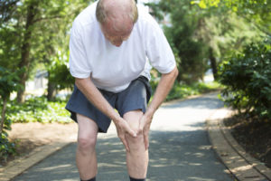
Before you get too far into your training, consider this; Four out of five amateur triathletes are injured while training, and three of those four are injured badly enough to be sidelined from their daily activities as well as their tri training.

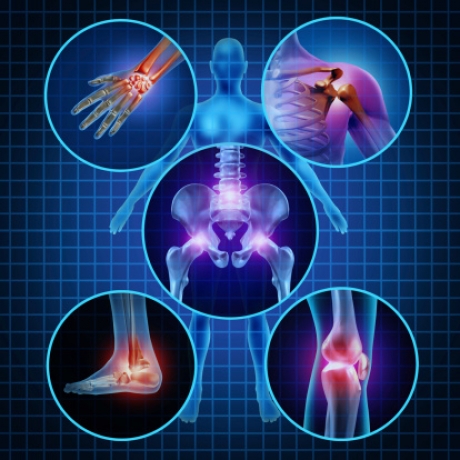
The complexity of the sport and the broad range of knowledge needed to train and compete safely are contributing factors. The triathlete must learn about appropriate equipment, proper body mechanics, nutrition and hydration, injury prevention and overall training programs that prepare the body for the stresses of a triathlon.


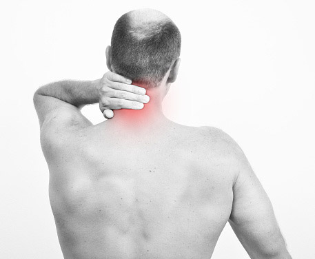
Cross training for three events – running, biking and swimming – also increases the risk of certain overuse injuries, and an untreated injury in one part of the body can lead to problems elsewhere. For example, a knee injury from running can cause extra stress on the back, leading to lower back pain when cycling; and the cumulative effect of swimming and cycling can fatigue calf muscles, making legs more susceptible to injury during a run. Improper training can be a vicious cycle, resulting in injury and, ultimately, derailing your resolution to compete in a triathlon.

The good news is, most non-traumatic injuries are related to training errors that can be corrected – and an orthopedic physician with specialized knowledge in sports medicine and injury prevention can help.

Train Smart

The best way to avoid traumatic injury is to undertake a training program that balances strength, flexibility and endurance through weight lifting, stretching and cross training. But training techniques are not one size fits all. Each individual has a unique combination of anatomy, strength, endurance and flexibility, so what works well for one athlete isn’t always the best route for another.

Understanding the relationships between musculoskeletal groups related to swimming, cycling and running is key to triathlon training. An orthopedic surgeon can evaluate your physical conditioning, analyze your training techniques and correct errors that can lead to potential injuries, as well as provide important input into a training program optimized for your needs.

Heal Smart

The best training program can’t prevent all injuries. When injuries do occur, there are three things you need to know:

  • How to evaluate the severity of the injury
  • How to treat an overuse injury
  • When to seek professional help

Congratulations for taking on a triathlon challenge. Whether you’re a first-time athlete or a seasoned tri gal or tri guy, training for and participating in the sport of triathlon involves a huge, months-long commitment. Unfortunately, sometimes things don’t go as planned and injury can overturn even the most dedicated competitor with the most detailed training plan.

When the unforeseen occurs, it is always best to seek medical evaluation from an experienced orthopedic physician. Often, if these issues are addressed early on, they can be treated without significant time loss from training and without surgery. An experienced orthopedic physician understands the sports medicine needs of patients and can be an important part of your team, helping you return to sport as soon as possible.

Preparing For A Triathlon

Make a plan:

  • Select a competition that’s right for you
  • Start your training gradually, begin with short distances and work up to longer distance, faster times and greater intensity
  • Consider recruiting a training buddy to help keep you on track with your goals
  • Check out websites such as USA Triathlon to locate training groups or routes in your area

Eat Properly

  • Consume foods rich in nutritional content and high in complex carbs
  • Increase your intake closer to race time, and supplement with energy gels and sports drinks as needed

Choose Proper Equipment

  • Research water temps to see if you need a wetsuit
  • Make sure your bike chains and pedals are working properly, and don’t forget your helmet
  • Wear comfortable training shoes and avoid sporting a new pair that may lead to blisters
  • If you experience severe or persistent pain while training, seek advice from an orthopedic physician before a more serious injury can occur

Bone and Joint Problems Associated with Diabetes

November is American Diabetes Month, a time set aside to raise awareness about diabetes and its associated risk factors.

Did you know that if you are an individual living with diabetes, you are at higher risk for some bone and joint disorders?

Certain factors such as nerve damage (diabetic neuropathy), arterial disease and obesity may contribute to these conditions, but often the cause isn’t clear.

If you feel that you are experiencing any of these symptoms, talk with your endocrinologist or consult a board certified orthopedic physician who will take your medical history of diabetes into account when diagnosing your condition.

Charcot Joint
Charcot (shahr-HOK) joint, also called neuropathic arthropathy, occurs when a joint deteriorates because of nerve damage – a common complication of diabetes. Charcot joint primarily affects the feet.
Symptoms include numbness and tingling or loss of sensation in the affected joints. They may become unstable, swollen or deformed. If detected early, progression of the disease can be slowed. Limited weight bearing activities and use of orthotic supports to the affected joint and surrounding structures can help.

Diabetic Hand Syndrome

Diabetic hand syndrome, also called cheiroarthropathy, is a disorder in which the skin on the hands becomes waxy and thickened. Eventually finger movement is limited. What causes diabetic hand syndrome isn’t known but it is most common in people who have had diabetes for a long time.
Over time, individuals with diabetic hand syndrome become unable to fully extend their fingers or press their palms together flat. Better management of blood glucose levels and physical therapy can slow the progress of this condition.

Osteoporosis
Osteoporosis is a disorder that causes bones to become weak and prone to fracture. People who have type 1 diabetes have an increased risk of osteoporosis.
Osteoporosis rarely causes symptoms in the early stages. Eventually, when the disease is more advanced, individuals can experience loss of height, stopped posture or bone fractures. A healthy lifestyle, including weight bearing exercise such as walking, and eating a well-balanced diet rich in calcium and vitamin D – including supplements if needed – are the best ways to address this condition.

Osteoarthritis
Osteoarthritis is a joint disorder characterized by the breakdown of joint cartilage. It may affect any joint in the body. People who have type 2 diabetes have an increased risk of osteoarthritis, likely due to obesity – a risk factor for type 2 diabetes – rather than to the diabetes itself.
Osteoarthritis may cause joint pain, swelling and stiffness as well as loss of joint flexibility or movement. Treatment involves exercising and maintaining a healthy weight, caring for and resting the affected joint, pain medication and, in some cases, surgery. Complimentary treatment such as acupuncture and massage can be helpful.

DISH
Diffuse idiopathic skeletal hyperostosis (DISH), also called Forestier disease, is a hardening of tendons and ligaments that commonly affects the spine. DISH may be associated with type 2 diabetes, perhaps due to insulin or insulin-like growth factors that promote new bone growth.
Affected individuals may experience pain, stiffness or decreased range of motion in any affected part of the body. Treatment involves managing symptoms, usually with pain medication, and in rare cases may require surgery to remove bone that has grown due to the condition.

Dupuytren Contracture
Dupuytren contracture is a deformity in which one or more fingers are bent toward the palm. It’s caused by thickening and scarring of connective tissue in the palm of the hand and in the fingers. This condition is common in people who have had diabetes for a long time.
People affected by dupuytren contracture may notice thickening of the skin on the palm of their hand. Eventually, they may not be able to fully straighten one or more fingers. Steroid injections may help reduce inflammation. Surgery, injections and a minimally invasive procedure called aponeurotomy to break apart the thick tissue are other options if the condition prevents the ability to grasp objects.

Frozen Shoulder
Frozen shoulder is a condition characterized by shoulder pain and limited range of motion. It typically affects only one shoulder. Although the cause is unknown, diabetes is a common risk factor.
Frozen shoulder causes pain or tenderness with shoulder movement, stiffness of the joint and decreased range of motion. If started early, aggressive physical therapy can help preserve movement and range of motion in the joint.

Carpal Tunnel Syndrome
Carpal tunnel syndrome is caused by a constriction of the hand’s central nerve by a ligament that runs across the palm. Diabetes may account for between 5 and 16 percent of all cases of carpal tunnel syndrome. The link between diabetes and carpal tunnel syndrome may be that the ligament becomes thickened in response to collagen glycation so that it presses on the nerve. Another possibility is that diabetic neuropathy – nerve disease – damages the nerves in the hand, making them more susceptible to carpal tunnel syndrome.
Strengthening and stretching exercises under the direction of a physical therapist may be helpful. Steroids and a wrist splint can be an interim measure, and surgery may be needed eventually.

PROTECT YOUR YOUNG ATHLETE FROM ORTHOPEDIC INJURY

Teen sports are great: They promote teamwork, jump-start a lifelong exercise habit, and provide an antidote to obesity. But teen athletes can also get hurt, which means they—and their parents and coaches—should be vigilant about prevention.

Sports injuries fall into two categories. Acute injuries, like a sprained ankle or torn ACL, occur suddenly, after a missed step or a midfield collision. Overuse injuries are caused over time by repetitive motion. Overuse injuries used to be fairly rare among teens and kids but increasingly, orthopedic specialists see teens with overuse injuries that used to plague mostly collegiate or pro athletes.

These injuries include damaged ulnar collateral ligament in the elbow (common in baseball pitchers, it can be fixed with so-called Tommy John reconstruction surgery), or osteochondritis dissecans, an overuse problem most commonly found in the knee that can result in loose bone or cartilage fragments in the joint.

One culprit: America’s youth sports culture. Immersion in high school teams, private club teams, traveling teams and sports summer camps mean more injuries. Many kids now specialize early and pursue a single sport through adolescence, rather than switching sports with the season. When young athletes do that, they lose the benefit of cross-training. Focusing on all-around athleticism keeps the body balanced and less vulnerable to injury.


There are ways to protect against both overuse and acute injuries. Proper conditioning is crucial. Young athletes new to sports should start by getting in good overall shape—including working on aerobic fitness, strength, and flexibility.

Start by being active for 30 to 60 minutes most days of the week, for at least six to eight weeks, before they get into sports. If not, it’s an absolute setup for injury. On the other hand, serious teen athletes may need to build more recovery time into their schedule, training hard on some days, but going easier and working on recovery and technique on alternate days.

Technique is important. Many young pitchers, for example, improperly rely on their arms for power rather than their trunk and legs. Done correctly, both strength training and working on the core muscles of the back and abdomen may prevent injury and boost performance. Sports-specific warm-up programs can also help.

When an injury does occur, its severity can be lessened by rest, prompt treatment and physical therapy. The incorrect reaction is to discourage teens from participating in sports. The risk of injury is far outweighed by the benefits of physical activity.

OVERUSE INJURIES IN YOUNG ATHLETES

Sports participation promotes the physical and emotional well-being of children, and also encourages a lifelong habit of exercise. Although the benefits of athletic activity are significant, too much activity can lead to injury.

In recent years, orthopedic physicians have begun to see young athletes with significant increase in overuse injuries. In most cases, these are sports related.

Overuse injuries occur gradually over time, when an athletic activity is repeated so often that some areas of the body do not have enough time to recover between playing. For example, overhand pitching in baseball can result in injuries to the elbow, and swimming is often associated with injuries of the shoulder.

Because young athletes are still growing, they are at greater risk of injury than adults. The consequences of overdoing a sport can include injuries that impair growth and may lead to long-term health problems.


When a young athlete repeatedly complains of pain, a period of rest from the sport is necessary. If pain persists, it is important to seek proper medical treatment. To ensure the best possible recovery, athletes, coaches and parents must follow safe guidelines to plan a return to the game.

Overuse injuries occur in a wide range of sports, from baseball and basketball to track, soccer and gymnastics. Some of these injuries are unique to a certain sport, such as throwing injuries of the elbow and shoulder that are prevalent in baseball players. The most common overuse injuries involve the knee and foot.

Overuse injuries can affect muscles, ligaments, tendons, bones and growth plates. In children, these structures are still growing, and the growth is generally uneven. Bones grow first, which pull at tight muscles and tendons. This uneven growth pattern makes young athletes more susceptible to muscle, tendon and growth plate injuries.

Growth plates are those areas of developing cartilage where bone growth occurs in children. The growth plates are weaker than the nearby ligaments and tendons. Repetitive stress can lead to injury of the growth plate and disrupt normal growth of the bone.

Concerned your young athlete may be developing an overuse injury? Make time to talk with a board certified orthopedic physician who specializes in the care of young athletes.

Rotator Cuff Injuries & Care

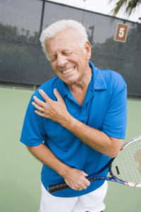
Rotator cuff tendonitis occurs when the tendons and muscles that help move the shoulder joint are inflamed or irritated. This condition commonly occurs in people who play sports that frequently require extending the arm over the head, such as tennis, swimming and pitching. Most people with rotator cuff tendinitis can regain full function of the shoulder without any pain after treatment.

Rotator cuff tendonitis affects the tendons and muscles that help move the shoulder joint. If you have tendinitis, it means that your tendons are inflamed or irritated. Rotator cuff tendinitis is also called impingement syndrome.

This condition usually occurs over time. It can be the result of keeping the shoulder in one position for a while, sleeping on the shoulder every night, or participating in activities that require extending the arm over the head. Sometimes, rotator cuff tendinitis can occur without any known cause.

Rotator Cuff Symptoms Worsen Over Time

The symptoms of rotator cuff tendinitis tend to get worse over time. Initial symptoms may be relieved with rest, but the symptoms can later become constant. Symptoms of rotator cuff tendinitis include:

  • Pain triggered by raising or lowering the arm
  • A clicking sound when raising the arm
  • Stiffness
  • Pain and swelling in the front of the shoulder and side of the arm
  • Pain that causes you to wake from sleep
  • Pain when reaching behind the back
  • A loss of mobility and strength in the affected arm

How Is Rotator Cuff Tendinitis Diagnosed?

Your orthopedic physician will begin by examining your shoulder to see where you’re feeling pain and tenderness. Your doctor will also test your range of motion and the strength of your shoulder joint. They may also examine your neck to check for conditions such as a pinched nerve or arthritis that can cause symptoms similar to rotator cuff tendinitis.

Your orthopedic physician may order imaging tests to confirm the diagnosis of rotator cuff tendinitis and rule out any other causes of your symptoms. An X-ray may be ordered to see if you have a bone spur. Your doctor may order an ultrasound or MRI to check for inflammation in the rotator cuff and to check for any tearing.

How Is Rotator Cuff Tendinitis Treated?

Initial treatment of rotator cuff tendinitis involves managing pain and swelling to promote healing. This can be done by:

  • Avoiding activities that cause pain
  • Applying cold packs to your shoulder three to four times per day
  • Taking nonsteroidal anti-inflammatory drugs (NSAIDs) like ibuprofen and naproxen
  • Additional treatment may include physical therapy and steroids

If nonsurgical treatment isn’t successful, your orthopedic physician may recommend surgery. Most people experience full recovery after having rotator cuff surgery. The most noninvasive form of shoulder surgery is accomplished via arthroscopy. This involves two or three small cuts around the shoulder, through which your surgeon will insert various instruments. One of these instruments will have a camera, so your surgeon can view the damaged tissue through the small incisions.

Home Care for Your Shoulder

There are several things you can do to help reduce pain from rotator cuff tendinitis. These techniques can also help prevent rotator cuff tendinitis or another flare-up of pain.

Shoulder self-care includes:

  • Using good posture while sitting
  • Avoiding lifting your arms repetitively over your head
  • Taking breaks from repetitive activities
  • Avoiding sleeping on the same side every night
  • Avoiding carrying a bag on only one shoulder
  • Carrying things close to your body

Read 788 times

Men and Osteoporosis

Osteoporosis is a disease that causes the skeleton to weaken and the bones to break. While most people think of osteoporosis as a woman’s disease, it poses a significant threat to millions of men in the United States.
There are many lifestyle habits that put men at increased risk of osteoporosis, yet few men recognize the disease as a significant threat to their mobility and independence.
Osteoporosis is called a “silent disease” because it progresses without symptoms until a fracture occurs. It develops less often in men than in women because men have larger skeletons, their bone loss starts later and progresses more slowly, and they have no period of rapid hormonal change and bone loss. However, in the past few years the problem of osteoporosis in men has become more recognized, particularly in light of estimates that the number of men above the age of 70 will continue to increase as life expectancy continues to rise.
Bone is constantly changing. Old bone is removed and replaced by new bone. During childhood, more bone is produced than removed, so the skeleton grows in both size and strength. For most people, bone mass peaks during the third decade of life. By this age, men typically have accumulated more bone mass than women. After this point, the amount of bone in the skeleton typically begins to decline slowly as removal of old bone exceeds formation of new bone.

Men in their fifties do not experience the rapid loss of bone mass that women do in the years following menopause. But by age 65 or 70, men and women are losing bone mass at the same rate, and the absorption of calcium, an essential nutrient for bone health throughout life, decreases in both sexes. Excessive bone loss causes bone to become fragile and more likely to fracture.
Fractures resulting from osteoporosis most commonly occur in the hip, spine, and wrist, and can be permanently disabling. Hip fractures are especially dangerous. Perhaps because such fractures tend to occur at older ages in men than in women, men who sustain hip fractures are more likely than women to die from complications.
In addition to age-related bone loss, there are many lifestyle behaviors, diseases and medications that can hasten bone loss in men. These include:
• Chronic diseases that affect the kidneys, lungs, stomach, and intestines, or alter hormone level
• Regular use of certain medications, such as glucocorticoids
• Undiagnosed low levels of the sex hormone testosterone
• Unhealthy lifestyle habits: smoking, excessive alcohol use, low calcium intake, and inadequate physical exercise
• Age. The older you are, the greater your risk
• Race. Caucasian men appear to be at particularly high risk, but all men can develop this disease.
Osteoporosis can be effectively treated if it is detected before significant bone loss has occurred. An orthopedic physician can order a medical workup to diagnose osteoporosis, including a complete medical history, x-rays, and urine and blood tests. The doctor may also order a bone mineral density test to determine your risk for fractures (broken bones), and measure your response to osteoporosis treatment.
In men, osteoporosis is often not diagnosed until a fracture occurs or a man complains of back pain and sees his doctor. This makes it especially important for men to inform their doctors about risk factors for developing osteoporosis, loss of height or change in posture, a fracture, or sudden back pain.
Once a man has been diagnosed with osteoporosis, his orthopedic physician may prescribe medications as well as a treatment plan including nutrition, exercise, and lifestyle guidelines for preventing bone loss.
Other possible prevention or treatment approaches include calcium and/or vitamin D supplements and regular physical activity.
Experts agree that both men and women should take the following steps to preserve their bone health:
• Avoid smoking, reduce alcohol intake, and increase physical activity
• Ensure a daily calcium intake that is adequate for your age
• Ensure an adequate intake of vitamin D
• Engage in a regular regimen of weight-bearing exercises in which bones and muscles work against gravity. This might include walking, jogging, racquet sports, climbing stairs, team sports, weight training, and using resistance machines
• Discuss with your doctor the use of medications that are known to cause bone loss, such as glucocorticoids
• Recognize and seek treatment for any underlying medical conditions that affect bone health

10 Ways to Build Healthy Bones and Keep Them Strong

Weak bones may seem like a problem of aging, but there’s plenty we can do early in life – in our 20s and even teens – to make sure bones stay healthy later in life.

Bones are the support system of the body, so it’s important to keep them strong and healthy. Bones are continuously being broken down and rebuilt in tiny amounts. Before about age 30, when bones typically reach peak bone mass, the body is creating new bone faster, but after age 30, bone building shifts and more bone is lost than gained.

Some people have a lot of savings in their “bone bank” because of factors including genetics, diet, and how much bone they built up as teenagers. The natural depletion of bone doesn’t affect these individuals too drastically. But in those with a smaller bone bank, when the body can’t create new bone as fast as the old bone is lost, osteoporosis can set in, causing bones to become weak and brittle and to fracture more easily. The disease is most common in postmenopausal women over the age of 65, and in men over the age of 70.

Although menopause and older age may seem like it is a long way off, once these milestones set in, it’s extremely hard to reverse. Since there’s no way of being 100 percent positive you’ll develop osteoporosis, the best way to counteract it is to take steps earlier in life to beef up bone mass (and prevent its loss) as much as possible.

Unfortunately, some are more likely than others to develop osteoporosis and weak bones in general, especially white and Asian postmenopausal women. But there are some things that can be changed to bump up bone mass. Here are 10 tips to make deposits in your bone bank for a healthier future.

1. Know your family history. Family history is a key indicator of bone health. Those with a parent or sibling who has or had osteoporosis are more likely to develop it.

2. Boost calcium consumption. When most people think bones, they think calcium. This mineral is essential for the proper development of teeth and bones. Calcium also contributes to proper muscle function, nerve signaling, hormone secretion, and blood pressure.

Help your body absorb calcium by pairing calcium-rich foods with those high in vitamin D. Foods that are good sources of calcium include yogurt, cheese, milk, spinach, and collard greens.

3. Don’t forget the vitamin D. Where there’s calcium, there must be vitamin D. The two work together to help the body absorb bone-boosting calcium. Boost vitamin D consumption by munching on shrimp, fortified foods like cereal and orange juice, sardines, eggs (in the yolks) and tuna, or opt for a vitamin D supplement.

4. Boost bone density with vitamin K. Vitamin K is mostly known for helping with blood clotting, but it also helps the body make proteins for healthy bones. Foods like kale, broccoli, Swiss chard, and spinach are high in vitamin K.

5. Pump up the potassium. Potassium isn’t necessarily known for aiding bone health: it’s a mineral that helps nerves and muscles communicate and also helps cells remove waste. But it turns out potassium may neutralize acids that remove calcium from the body.

Studies in both pre- and postmenopausal women have shown that a diet high in potassium can improve bone health. Load up on potassium by eating foods sweet potatoes, white potatoes (with the skin on), yogurt, and bananas.

6. Make exercise a priority. Regular exercise is key to keep a number of health issues at bay, and bone health is no exception. Living a sedentary lifestyle is considered a risk factor for osteoporosis.   What type of exercise is most effective? Weight-bearing exercises like running, walking, jumping rope, skiing, and stair climbing keep bones strongest. Bonus for the older readers: improved strength and balance helps prevent falls (and the associated fractures) in those who already have osteoporosis.

7. Consume less caffeine. Caffeine does have some health benefits, but unfortunately not for our bones. Too much of it can interfere with the body’s ability to absorb calcium.

8. Cool it on the booze. But like caffeine, there’s no need to quit entirely. While heavy alcohol consumption can cause bone loss (because it interferes with vitamin D doing its job), moderate consumption (that’s one drink per day for women, two per day for men) is fine — and recent studies actually show it may help slow bone loss.

9. Quit smoking. Here’s yet another reason to lose the cigarettes: multiple studies have shown that smoking can prevent the body from efficiently absorbing calcium, decreasing bone mass.

10. Don’t be an astronaut. Not to squash any childhood dreams, but because of hours and hours of weightlessness and low-calcium diets, astronauts often suffer from space-induced osteoporosis. Space-anything sounds kind of awesome, but space bones definitely aren’t: astronauts can lose up to 1 to 2 percent of their bone mass per month on a mission! For those who simply must visit the moon, there is a possible solution: two studies have found that vitamin K can help build back astronauts’ lost bone — more than calcium and vitamin D.

Orthopedic Injuries Rise with Hoverboard Popularity

Hoverboards, one of the hottest gifts for both kids and adults this past holiday season, are also becoming a popular reason for visits to the emergency room.
An item that is a mixture of a skateboard and a Segway, a hoverboard allows the rider to stand on a platform that will accelerate and move freely in the direction of the rider’s weight. While fun and perhaps exhilarating, hoverboards are also putting some riders at risk.
Orthopedic physicians are seeing an uptick in injuries to bones and joints, wrist and ankle injuries, and fractures. Other injuries associated with hoverboards include spinal fractures, facial lacerations, head trauma with concussion, and contusions. Some injuries are more serious because of the height and speed behind the fall.
Riders need to have good balance on the devices, which don’t have a handle. The faster riders go, the higher the injury risk. Hoverboards can speed up to 12 miles per hour.
To avoid and prevent serious injury from hoverboards, follow these safety tips:
* Wear a helmet
* Wear wrist guards, elbow pads and knee pads
* Practice and become familiar with the hoverboard in an open, soft flat field
* Be mindful of vehicles and other forms of traffic and impediments (stay off the roads)
Supervise children using hoverboards and take the same precautions as you would for using a scooter, bicycle or roller blades, especially wearing a certified safety helmet.
When it comes to hoverboard safety for children and adults alike, start slow and make sure you have something stable to hold on to while you’re getting a feel for the hoverboard.

Don’t Delay Treatment for Winter Ankle Injuries

Icy, snowy conditions can pose serious health and safety threats.

Over the winter months, we see a definite increase in fractures (breaks in the bones), sprains and other orthopedic injuries, resulting from slips and falls, and even from skidding accidents involving motor vehicles.

Broken ankles are one of the most common fracture types and can be caused by slipping and twisting the ankle while trying to navigate slippery surfaces.

If you fall on the ice and hurt your ankle this winter, don’t put off waiting to see a doctor. The ability to walk or hobble on your injured ankle doesn’t necessarily mean that your ankle isn’t broken or badly sprained. It’s best to have an injured ankle evaluated by an orthopedic surgeon as soon as possible. Only an x-ray and thorough examination by an orthopedic surgeon can determine the extent of your injury.

Putting weight on an injured ankle joint can worsen the problem and may lead to chronic instability, joint pain and even arthritis later in life. If you can’t see an orthopedic surgeon or visit an emergency room right away, follow the R.I.C.E. method – Rest, Ice, Compression and Elevation – until medical care is available.

The ankle joint is especially vulnerable to serious injury from hard falls on ice. Ice accelerates the fall and often causes more severe trauma, because the foot can go in any direction after it slips as well as bearing the weight of your body as you fall.

Falling on winter ice can cause simultaneous ankle sprains and fractures. It is possible to both fracture and sprain an ankle from a fall, and a bad sprain can mask the fracture.

Most ankle fractures and some sprains are treated by immobilizing the joint in a cast or splint.  Surgery may be needed to repair fractures with significant misalignments. New surgical plates and screws enable orthopedic surgeons to repair these injuries more quickly and with less surgical trauma.

Newer bone-fixation methods require smaller incisions, minimizing tissue damage and bleeding, reducing the risk of infection, and accelerating the healing process.

Lifestyle Changes To Manage Arthritis Pain

What you do or don’t do every day can have a major impact on your joints and the level of arthritis pain you experience. Arthritis is a leading cause of pain and disability worldwide.

Whatever your condition, you can work to stay ahead of your pain by:

  • Talking to your orthopedic physician about your symptoms, arthritis related or not. Sometimes seemingly unrelated problems are connected. Your doctor will know for sure.
  • Give your orthopedic physician complete information about your medical condition and medications, including over the counter medications and supplements.
  • Ask your orthopedic physician for a clear definition of the type of arthritic you have.
  • Find out whether any of your joints are already damaged.

Arthritis affects about 36 percent of obese American adults, according to the Centers for Disease and Prevention. A healthy body weight is important to your body’s overall health, including your joints. Joints that are stressed by bearing the burden of excess pounds show more wear and tear, inflammation and stiffness, leading to arthritic pain.

Consider making these changes to help reduce your arthritis pain:

  • Exercise for at least 20 minutes every day to keep the pounds off and reduce arthritis pain and stiffness.
  • Eat a healthy diet of low-fat, low-calorie foods and plan your meals and snacks to be sure you are getting lots of fruits and vegetables through your diet.
  • Get plenty of vitamin C to help manage joint inflammation. Studies have shown that vitamin C may be helpful in managing inflammation, so dig into an orange or have a glass of grapefruit juice.
  • Manage your weight. Even a few extra pounds can worsen your arthritis pain, so talk to your doctor to determine your healthiest weight. Focus on that number as your goal, and track your weight loss until you reach it.
  • Quit smoking. Smoking causes stress on connective tissues, which leads to more arthritis pain.
  • Avoid alcohol. Don’t medicate yourself with alcohol to manage pain; it will only create more problems and add calories to your diet.

Schedule an appointment with a board certified orthopedic physician to get tips on exercise and other common concerns when coping with arthritis symptoms and arthritis pain.